


Product Overview
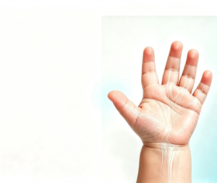
Postaxial polydactyly is a congenital hand difference in which an additional digit develops on the ulnar side of the hand, next to the small finger. The condition ranges from a small, rudimentary digit attached by a narrow soft-tissue pedicle to a more developed duplicated digit with skeletal and joint connections.
This clinical portal serves pediatric hand surgeons, orthopedic departments, reconstructive plastic surgery teams, and healthcare institutions. We provide structured case reviews, surgical planning, and secondary reconstructive strategies for pediatric postaxial polydactyly (ulnar polydactyly).
Anatomical Classification & Decision Matrix: Type A vs. Type B
|
Feature |
Type A - Fully Formed / Skeletal |
Type B - Rudimentary / Pedunculated |
|
Anatomical Structure |
Contains more developed phalangeal structures and may articulate with the fifth metacarpal or a duplicated metacarpal. |
Typically presents as a rudimentary soft-tissue appendage connected by a narrow pedicle that may contain neurovascular structures. |
|
Radiographic Assessment |
Radiographs are commonly used to define phalangeal, metacarpal, joint, and skeletal attachment anatomy. |
Imaging may not be necessary when the digit is clearly soft-tissue based; it may be considered when a bony or cartilaginous connection is suspected. |
|
Primary Clinical Concerns |
Joint instability, angular deformity, residual malalignment, and tendon or soft-tissue imbalance may require consideration. |
Potential neuroma symptoms, residual nubbin or contour irregularity, wound complications, and incomplete tissue removal may require consideration. |
|
Reconstructive Focus |
May include preservation or reconstruction of joint stability, collateral ligament structures, tendon balance, and alignment after removal of the less functional component. |
Focuses on controlled removal or ligation of the accessory digit, with appropriate management of the digital nerve, vascular structures, and soft-tissue contour. |
|
Typical Treatment Setting |
Treatment may require an operating-room setting with an anesthesia plan appropriate for the child's age and procedure. |
Selected cases may be managed in an outpatient or minor-procedure setting, depending on the child's age, anatomy, treatment technique, and clinical environment. |
|
Treatment Planning Priority |
Preserve useful thumb-side hand function, maintain stability, and address skeletal and soft-tissue abnormalities where necessary. |
Achieve safe removal while minimizing residual tissue, contour irregularity, and symptomatic nerve-related complications. |
Surgical Excision vs. Suture Ligation for Type B Polydactyly
|
Clinical Parameter |
Direct Surgical Excision |
Suture / Clip Ligation |
|
Anatomical Control |
Allows direct visualization of the accessory digit, pedicle, and relevant neurovascular structures during removal. |
Relies on ligation or clipping of the pedicle, followed by ischemic separation of the accessory digit. |
|
Nerve Management |
The digital nerve can be identified and managed directly according to the surgeon's technique and the individual anatomy. |
The nerve may remain within or near the ligated pedicle, which may contribute to symptomatic neuroma or hypersensitivity in some cases. |
|
Contour & Residual Tissue |
Direct excision allows controlled removal of excess tissue and primary wound closure, which may provide a more predictable residual contour. |
Separation of the accessory digit may leave a residual nubbin, scar, or contour irregularity in some patients. |
|
Wound Management |
The wound is closed under direct visualization, allowing the surgeon to manage skin edges and soft-tissue contour at the time of treatment. |
The treated tissue separates over time, so wound appearance develops during the healing process. |
|
Potential Complications |
Possible complications include infection, bleeding, scarring, sensory symptoms, or residual deformity, although the risk varies by case. |
Reported concerns include infection, incomplete separation, residual tissue, scar or contour irregularity, and possible secondary treatment. |
|
Revision Considerations |
Direct removal may allow the surgeon to address the pedicle and surrounding tissue during the initial procedure. |
Residual nubbin, symptomatic scar, or other persistent tissue may require subsequent evaluation or revision in selected cases. |
|
Typical Treatment Setting |
May be performed in an outpatient or operating-room setting depending on the child's age, anesthesia requirements, and clinical circumstances. |
May be considered in selected pediatric or newborn settings, depending on the pedicle anatomy, age, and treating clinician's protocol. |
|
Clinical Decision |
Often considered when precise anatomical control, nerve management, and soft-tissue contour are priorities. |
May be considered for selected Type B cases when the anatomy and clinical setting are appropriate. |
Advanced Surgical Protocols for Complex Type A Reconstruction
In Type A cases, simple ablation of the extra digit is insufficient and frequently leads to ulnar instability of the small finger MCP joint. Our surgical protocol incorporates three core reconstructive steps:
1. Ulnar Collateral Ligament (UCL) Preservation and Anchoring
When the duplicated outer digit is excised, the ulnar collateral ligament of the metacarpophalangeal (MCP) or interphalangeal (IP) joint is preserved with a periosteal cuff. It is tensioned and re-anchored to the retained fifth metacarpal head using suture anchors or transosseous sutures to ensure lateral joint stability under grip load.
2. Tendon Realignment and Insertion Centering
Extensor digiti minimi (EDM) and flexor digitorum profundus (FDP) slips insertion points are frequently eccentric. Surgeons must release anomalous abductor slips, centralize the functional flexor and extensor mechanisms over the axial line of the retained digit, and perform intrinsic muscle rebalancing where required.
3. Metacarpal Resection and Corrective Osteotomy
In broad-based duplications with bifurcated or Y-shaped fifth metacarpals, a longitudinal or closing-wedge osteotomy is executed to remove the extra articular facet. Internal stabilization is achieved using smooth, longitudinal Kirschner wires (K-wires) positioned to protect the epiphyseal growth plates.
Revision Surgery for Post-Treatment Secondary Deformities
Our center frequently handles secondary procedures for patients with unsatisfactory primary outcomes. Common revision indications include:
- Symptomatic Neuromas: Secondary to bedside ligation where the digital nerve was entrapped in scar tissue. Treatment involves scar release, neuroma resection, and deep muscular transposition.
- Ulnar Deviation / Joint Instability: Resulting from unaddressed ligamentous deficiency following primary Type A excision. Managed via dynamic tendon transfer and collateral ligament reconstruction.
- Residual Bony/Soft-Tissue Prominences: Requiring secondary contouring and scar Z-plasty.
Standardized Case Submission Protocol for Institutional Partners
To streamline clinical case reviews and preliminary surgical consultations, partner institutions are requested to gather the following parameters prior to submission:
1. Patient Profile: Current age in months, sex, affected side (Right, Left, Bilateral), and presence of associated congenital syndromes.
2. Clinical Photography:
- Static dorsal and volar views of both hands in neutral position.
- High-resolution close-up of the ulnar border showing the pedicle width or joint base.
3. Radiographic Imaging: Standard AP and oblique hand X-rays in DICOM or high-resolution JPEG format.
4. Prior Intervention Data: Operational notes, date of procedure, and clinical history if evaluating a revision case.
5. Primary Consultation Goal: Assessment for primary reconstruction, residual tissue excision, scar revision, or joint stabilization.
FAQ
Q: What is the recommended age for Type A postaxial polydactyly reconstruction?
A: Reconstruction is typically scheduled between 9 and 18 months of age. This timing provides adequate anatomical size for delicate ligamentous and capsular reconstruction while establishing structural stability prior to the development of fine motor pinch and grasp habits.
Q: How is digital nerve involvement handled during Type B digit removal?
A: To prevent painful post-operative neuromas, the digital nerve within the pedicle must be identified under magnification, gently pulled down under light tension, sharply transected, and allowed to retract deep into the surrounding hypothenar soft tissue away from the skin incision.
Q: Is plain radiography required for every Type B case?
A: Radiographs are not strictly mandatory for narrow, fully pedunculated soft-tissue tags lacking palpable cartilage. However, if any firmness, broad base, or uncertainty regarding skeletal involvement exists at the base, X-rays are recommended prior to intervention.
Q: What is the post-operative recovery protocol for Type A reconstructive surgery?
A: Following osteotomy or joint reconstruction, the hand is immobilized in a long-arm cast or custom thermoplastic splint for 4 to 6 weeks. Any temporary smooth K-wires are removed in an outpatient clinic setting once radiographic bony union is confirmed.
Medical Disclaimer
The information provided on this page is intended for professional educational purposes and institutional case evaluation only. It does not replace an in-person medical diagnosis or clinical consultation. Surgical recommendations, timing, and outcomes depend on individual anatomical assessment and patient-specific factors.
Hot Tags: postaxial polydactyly treatment, China postaxial polydactyly treatment doctor


