


Product Overview
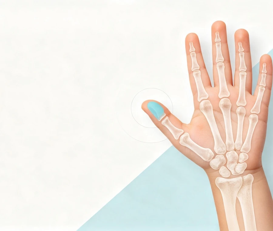
Pediatric preaxial polydactyly-commonly referred to as radial polydactyly or thumb duplication-is a congenital hand difference characterized by partial or complete duplication of the radial digit (thumb).In clinical hand surgery, treatment is rarely a simple matter of resecting the smaller or accessory digit. In the majority of presentations, the duplicated components share, divide, or maldistribute critical anatomical structures.
Led by International Surgical Expertise: Dr. Jianghai Chen
Dr. Jianghai Chen, MD, PhD
Associate Professor / Chief Physician of Hand Surgery
Wuhan Union Medical College Hospital
Dr. Jianghai Chen completed his MD in Surgery at Huazhong University of Science and Technology and earned his PhD at KU Leuven, Belgium. As a doctoral supervisor, editorial board member of international surgical journals, and leading hand surgeon, Dr. Chen specializes in pediatric microsurgery and complex congenital hand differences.
Preoperative Clinical Evaluation
Comprehensive surgical planning requires a multi-dimensional assessment combining clinical examination and static/dynamic imaging.
1. Physical Examination
- Alignment and Axis: Degree of radial or ulnar deviation (angulated deformities).
- Joint Stability: Passive and active stress testing of the MCP and IP joints to detect lateral laxity.
- Tendon Dynamics: Assessment of active flexion, extension, and eccentric tendon pulls caused by abnormal bifurcations.
- Soft Tissue and Nail Envelope: Measurement of relative thumb volume, nail bed widths, and first web-space skin deficiency.
2. Radiographic Assessment
Standard Radiographs (AP, Lateral, Oblique Views): Define the skeletal level of duplication, joint congruence, bone maturity, and the presence of abnormal ossification (e.g., trapezoidal or Delta phalanx).
High-Resolution Ultrasonography (When Indicated): Useful in infants prior to secondary ossification center appearance to evaluate unossified cartilage structures, joint surfaces, and soft-tissue alignments.
The Wassel Classification Framework
The Wassel classification serves as a universal baseline for communicating the anatomical level of duplication (Types I through VII):
|
Classification |
Anatomical Level |
Structural Description |
|
Wassel Type I |
Distal Phalanx |
Bifid distal phalanx sharing a single joint surface |
|
Wassel Type II |
Distal Phalanx |
Complete duplication of the distal phalanx |
|
Wassel Type III |
Proximal Phalanx |
Bifid proximal phalanx with duplicated distal phalanges |
|
Wassel Type IV |
Proximal Phalanx |
Most common pattern. Complete duplication of proximal phalanx on a shared metacarpal head |
|
Wassel Type V |
Metacarpal |
Bifid metacarpal structure |
|
Wassel Type VI |
Metacarpal |
Complete duplication of the metacarpal bone |
|
Wassel Type VII |
Triphalangeal / Complex |
Duplication associated with a triphalangeal thumb pattern or complex joint dysplasia |
Surgical Decision Workflow
Our clinical approach follows a structured, step-by-step evaluation framework:
- Initial Clinical Examination and Functional Assessment
- Radiographic and Ultrasound Anatomical Mapping
- Wassel Classification and Soft-Tissue Deficit Categorization
- Identification of Shared vs. Hypoplastic Tendons, Ligaments, and Joint Surfaces
- Selection of the Primary Functional Thumb Component
Execution of Reconstructive Strategy (Ablation with Reconstruction vs. Combined/Modified Procedures)
Key Reconstructive Techniques
1. Collateral Ligament Reconstruction & Joint Capsulorhaphy
When the accessory thumb is excised, its attached collateral ligament (typically the radial collateral ligament) must be preserved with a small periosteal flap and reanchored securely to the retained thumb's metacarpal head or phalanx using bone anchors or transosseous sutures. This prevents post-operative lateral joint instability.
2. Tendon Re-centering and Rebalancing
Duplicated thumbs often present with eccentric flexor or extensor tendon insertions that cause progressive zig-zag or angular deformities. Surgical planning involves detaching the anomalous tendon slip and re-centering its insertion onto the midline of the retained thumb's distal phalanx.
3. Corrective Osteotomy
If the retained thumb exhibits residual structural angulation greater than 10 to 15 degrees-or if a Delta phalanx is present-a closing or opening wedge osteotomy is performed and temporarily stabilized with smooth Kirschner wires (K-wires) to restore the longitudinal mechanical axis.
4. Modified Bilhaut-Cloquet Procedure
In select cases where both duplicated thumbs are severely hypoplastic and of similar size, a combined procedure may be considered. To avoid the classical complications of nail ridge deformities and stiff interphalangeal joints, modern surgical teams utilize a "modified Bilhaut-Cloquet" approach, combining skeletal and soft-tissue volume while preserving a single intact nail bed and joint capsule whenever possible.
Primary Surgical Goals and Long-Term Outcomes
Joint Stability: Secure MCP and IP joint stability to withstand opposition forces.
Axial Alignment: Elimination of angular deviation (zigzag collapse) under load.
Preserved Mobility: Maintenance of functional range of motion for precision pinch and power grip.
Cosmetic Harmony: Symmetrical thumb girth, length, and nail bed appearance relative to the contralateral thumb.
Physeal Protection: Meticulous preservation of growth plates (physes) to allow normal longitudinal growth throughout pediatric development.
Complex and Revision Cases
Secondary or revision cases often present following primary procedures performed elsewhere that resulted in residual radial/ulnar deviation, joint stiffness, or "zigzag" deformity (common in post-type IV reconstructions).
Revision surgical planning requires comprehensive reassessment to identify:
- Unaddressed tendon imbalance
- Unreconstructed lateral collateral ligament laxity
- Progressive physeal arrest or bony malalignment
Treatment often combines soft-tissue release, tendon re-routing, ligament reconstruction, and corrective osteotomies.
Information Required for Clinical Case Review
To facilitate professional case review and peer-to-peer clinical discussion, clinical teams and families are encouraged to provide the following information:
Patient Age: Exact age/month at evaluation
Laterality: Right, left, or bilateral involvement
Radiographs: Standard AP and Lateral X-rays clearly showing the hand and wrist bones
Clinical Photographs:
Dorsal view of both hands side-by-side
Volar (palmar) view
Dynamic grasp / opposition photographs or short video
Surgical History: Details of any previous procedures, including dates and operative notes
Primary Clinical Concerns: Functional limitation, joint weakness, angular deviation, or appearance
FAQ
Q: What is the ideal age for pediatric thumb duplication surgery?
A: Surgery is typically performed between 9 and 18 months of age. At this stage, skeletal and soft-tissue structures are larger, making reconstruction precise while allowing the child to develop normal pinch and grasp patterns without developmental delay.
Q: Will there be visible scarring after reconstruction?
A: Incisions are placed along natural skin creases or designed as zig-zag (Z-plasty) incisions to prevent straight-line scar contracture. Scar management protocols, including silicone sheeting and massage, begin after complete wound healing.
Q: How long is the hand immobilized after surgery?
A: When bone osteotomies or major ligament/tendon reconstructions are performed, the hand is immobilized in a long-arm cast or custom splint for 4 to 6 weeks. Any temporary K-wires are typically removed in the clinic setting at that time.
Q: Why is simple excision of the extra thumb insufficient?
A: The duplicated thumbs frequently share vital structures. Removing one thumb without re-attaching its collateral ligaments or re-balancing its tendons inevitably leads to joint instability, weakness, and progressive angular deformity over time.
Medical Disclaimer
The information provided on this page is intended for professional educational purposes and institutional case evaluation only. It does not replace an in-person medical diagnosis or clinical consultation. Surgical recommendations, timing, and outcomes depend on individual anatomical assessment and patient-specific factors.
Hot Tags: pediatric preaxial polydactyly surgery, China pediatric preaxial polydactyly surgery doctor


