


Would you believe it if I told you the two hands shown in the pictures actually belong to the same hand?

Left Hand: Before and After Syndactyly Surgery
This isn't a "magic trick" - it's the real before-and-after of a syndactyly surgery.
The child's mother happily shared a story with me:
Because the child previously had "diamond-shaped" syndactyly in both hands, many other children were curious about it. After the surgery and recovery, those same children saw her hands and were puzzled:
"Weren't this girl's fingers stuck together? How come they're fine now? Did I remember it wrong?"

Right Hand: Before and After Syndactyly Surgery
The little girl's recovery has been excellent. The scarring, tension lines, and mobility of both hands have greatly improved - not only making the parents happy but also bringing joy and satisfaction to us as doctors.
Originally, her middle and ring fingers on both hands were fused in a very complex way. The distal phalanges were completely joined, and there was a wide bony bridge between the middle phalanges, pushing the fingers apart. This made both hands appear in a diamond shape from the outside.
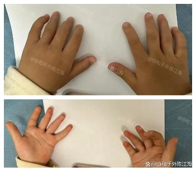
Post-Surgery
Such a complex form of syndactyly added considerable difficulty to the surgery.
Many people asked: How could even the fingernails be separated?
From the pictures, it's clear that the child has full nails, which seems "magical" to many - but in fact, with careful surgical planning, it's possible to divide the nail even in such cases.

Rare "Diamond-Shaped" Syndactyly
If you closely observe the nails at the fused area before surgery, they appear as one complete nail without a clear boundary. Apart from being slightly wide, they are not very different from the surrounding fingers.
During finger separation surgery, we split this nail "in two." The division itself is simple, but the problem arises afterward: the bone surface of the distal phalanx becomes exposed. At this stage, we need to find a way to encourage surrounding skin to gradually grow over it.
Since this involves the distal phalanx, it's difficult to use flaps to cover the area, and the success rate of skin grafting is relatively low. This type of wound is suitable for artificial dermis.

Post-Surgery
We use artificial dermis to cover the wound and create a relatively complete, normal-looking nail groove. Then, with some surrounding skin, we construct the nail edge and side walls, allowing the child to retain a natural-looking nail and improved finger function.
With the distal issue resolved, the proximal area is relatively easier to handle. Due to the child's unique anatomy, we had to consider in pre-op planning whether an osteotomy was needed. Fortunately, during surgery, we found that the proximal interphalangeal joints of the middle and ring fingers could be maintained in an extended position using Kirschner wires, so no osteotomy was necessary.
With internal fixation via Kirschner wires, external bracing, functional rehabilitation, and scar treatment, the child's hands are gradually growing and recovering well.
The surgery gave her a chance - and now the parents' role is to provide a safe and stable environment to support her recovery.